Core Imaging Instrumentation
Equipment
- FEI Quanta 200 Variable pressure Scanning Electron Microscope with Energy Dispersive X-Ray Microanalysis
- Zeiss LSM 700 Laser Scanning Fluorescence Inverted Confocal Compound Light Microscope
- Zeiss LSM 800 Laser Scanning Fluorescence Upright Confocal Compound Light Microscope
- Olympus BX-41 Compound Light Microscope with Phase and Fluorescence Imaging
- Olympus BX-40 Compound Light Microscope with Phase and Fluorescence Imaging
- Nikon Eclipse TE200 Inverted Compound Light Microscope with Phase Optics
- Olympus SZX10 Stereo Light Microscope with Oblique Lighting
- Leica MZ125 Stereo Light Microscope with Leica Camera IC80HD
- Zeiss Stemi 2000-C on a Boom-stand
- Zeiss Stemi 305 with integral camera and IPad Min
- Nikon D5100 Digital SLR camera, with 40 and 55 mm macro lenses and a Sigma EM-140 DG Macro Flash
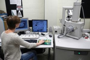
FEI Quanta 200 Variable pressure Scanning Electron Microscope with Energy Dispersive X-Ray Microanalysis
The SEM offers much greater depth of field, resolution, and much greater range of magnification (from about 40x to 50,000x) than light microscopy. The SEM images surface features. The low vacuum and ESEM modes of imaging require little or no specimen preparation thereby eliminating time and potential artifacts due to chemical fixation, drying and sputter coating. X-Ray Elemental Microanalysis is easy and quick even for a novice.

Zeiss LSM 700 Laser Scanning Fluorescence Inverted Confocal Compound Light Microscope
This microscope offers exceptional fluorescence imaging due to its Laser light sources and Confocal optics. Differential interference contrast is also offered. Solid State Lasers include 405nm, 635nm, 555nm and 488nm. See Dr. Ables (Biology Dept.) for permission to use this scope and for training.
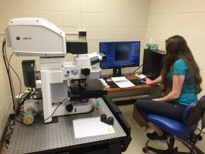
Zeiss LSM 800 Laser Scanning Fluorescence Upright Confocal Compound Light Microscope
This microscope offers exceptional fluorescence imaging due to its Laser light sources, GaAsP detection, and Confocal optics. Laser lines include 405nm, 488nm, 561nm and 640nm. Differential interference contrast is also offered. It also has 20 and 40x water immersion objectives. See Dr. Erickson (Biology Dept.) for permission to use this scope and for training.

Olympus BX-41 Compound Light Microscope with Phase and Fluorescence Imaging
The Olympus BX-41 is an easy to use, research grade compound light microscope with brightfield, darkfield, phase and reflective fluorescence optics (with DAPI, FITC and TRITC fluorescence cubes).

Olympus BX-40 Compound Light Microscope with Phase and Fluorescence Imaging
The Olympus BX-40 similar to the BX-41. With Rhodamine and FITC fluorescence cubes.
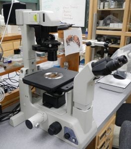
Nikon Eclipse TE200 Inverted Compound Light Microscope with Phase Opti
This microscope currently possesses 10x, 20x and 40x high-working distance phase objectives. It is also coupled with a 2x optivar.

Olympus SZX10 Stereo Light Microscope with Oblique Lighting
This high-magnification (6.3-94.5x) stereo microscope is ideal for fish embryos and other translucent subjects considering the optimized Oblique lighting.
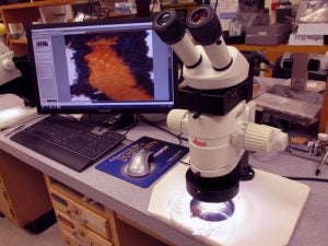
Leica MZ125 Stereo Light Microscope with Leica Camera IC80HD
This stereo microscope has total magnifications at the ocular lenses of 8-100x. Illumination is provided by an adjustable LED light source.
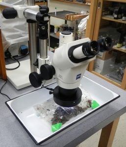
Zeiss Stemi 2000-C on a Boom-stand
This stereo scope on a boom-stand is ideal for situations requiring a large reach over, for example, a large pan for sorting specimens like insects, plants, and others. Magnification is from a low of 6.5x to 50x.

Zeiss Stemi 305 with integral camera and IPad Min
This stereo scope is reserved for educational purposes in a laboratory environment. The scope acts as a WiFi hub and can transmit pictures to student Ipads and Iphones in the laboratory or can be connected by HMDI through a Lightning Digital AV adapter to a large screen monitor.

Nikon D5100 Digital SLR camera, with 40 and 55 mm macro lenses and a Sigma EM-140 DG Macro Flash
SLR camera with a macro lens essentially spans the magnification range from what your eye observes to the low-end magnification range of a stereo microscope.